Hover to pan and click to magnify. Click again to pan at full screen.
Clinica Montallegro, Genova, IT, Clinical Research Department, Fidia Farmaceutici S.p.A., Abano Terme, Padova, IT
Hover to pan and click to magnify. Click again to pan at full screen.
Dr. Virginia Priano, Dr. Irene Beriotto, Dr. Nicola Giordan, Prof. Ferdinando Priano
Clinica Montallegro, Genova, IT, Clinical Research Department, Fidia Farmaceutici S.p.A., Abano Terme, Padova, IT
Background
Osteoarthritis (OA), is a disabling joint disorder characterized by articular cartilage degeneration and inflammation (Martel-Pelletier, 2016). Disease symptoms include pain, stiffness and impaired function with a resulting major negative impact on daily activities and patient’s quality of life. (Lane, 2011; Xie, 2016; Cross, 2014). At present, no curative therapy exists and treatment options are aimed at managing symptoms and restore function (Altman, 1998; Jordan, 2003; Zhang, 2007, 2008, 2010a; Mc Alindon et al., 2014).
Over the last two decades, therapies involving intra-articular injection of autologous cell-derived preparations have gained increasing attention (Di Matteo, 2019; Ha, 2019). Intra-articular administration of those preparations aims to enrich the degenerated cartilage with mesenchymal cells possibly able to differentiating into tissue-specific, regenerating cell populations and/or secrete anti-inflammatory and pro-regenerative growth factors able to interact with the tissue microenvironment (Shi, 2017; Delanois, 2019). Adipose-derived stem/stromal cells (ADSCs) have been widely investigated as source of mesenchymal stem cells (MSCs) (Bora, 2017; Ranmuthu, 2018; Di Matteo, 2019b). When extracted from adipose tissue by enzymatic digestion, they are contained in an aqueous fraction, known as the stromal vascular fraction (SVF), which contains endothelial precursor and differentiated cells, macrophages, smooth muscle cells, lymphocytes, pericytes, pre-adipocytes and growth factors (Nguyen, 2016; Guo, 2016; Bora, 2017; Pak, 2018). Both ADSCs and SVF have been used in the field of regenerative and reconstructive medicine, with some preliminary evidences of the superiority of SVF (Bora, 2017). Intra-articular injection of adipose tissue has been proven to be effective and safe for the treatment of knee OA in a series of clinical trials (Fodor, 2015; Arthurs, 2018; Cattaneo, 2018; Hudetz, 2017; Kim, 2016; Russo, 2017). Traditional separation of SVF from adipose tissue requires extensive tissue manipulation (enzymatic digestion, cell expansion) and dedicated facilities and personnel, thus limiting the use of SVF in the clinical setting (Bora, 2017; Pak, 2018). A system for the preparation of adipose tissue enriched in SVF (at-SVF) within a single surgical session, enzyme-free and with minimal manipulation has been recently developed (Hy-tissue SVF Separation System, Fidia Farmaceutici S.p.A., Abano Terme, Italy). At present, evidence concerning at-SVF effectiveness is limited to a randomized controlled clinical trial with a 6-month follow-up for the treatment of Achilles tendinopathy (Usuelli, 2018).
The present retrospective study aims to assess the efficacy and safety of a single intra-articular injection of at-SVF, prepared using the Hy-tissue SVF system, for the management knee OA symptoms over a 6-month period.
Methods
Data from 25 subsequent patients aged 25 to 85 years, with a documented diagnosis of knee OA K-L grade II-III, who received an intra-articular injection of at-SVF were collected in a suitable database and analysed. Clinical assessment were performed at 1, 3 and 6 months after treatment according the center’s standard clinical practice. The investigation involved a single center in Italy (Clinica Montallegro, Genova, IT). The study was conducted in compliance with the ethical principles of the Declaration of Helsinki, Good Clinical Practices International Conference on Harmonisation (ICH) Guidelines as well as the principles outlined in the European Regulation UE 2016/679 (GDPR) concerning protection of personal data.
Study Objectives
The primary objective of the present retrospective observational study was to evaluate the benefits of intra-articular injection of adipose tissue enriched in SVF on pain due to knee osteoarthritis (OA) up to 6 months (WOMAC pain (A) subscale).
Secondary objectives were:
• To evaluate the benefits of intra-articular injection of adipose tissue enriched in SVF on knee OA stiffness and physical function and overall benefits up to 6 months (WOMAC stiffness (B) subscale, WOMAC physical function (C) subscale, Total WOMAC Index);
• To evaluate the local tolerability and safety of intra-articular injection of adipose tissue enriched in SVF up to 6 months.
Results
Complete baseline patient characteristics are summarized in Table 1.
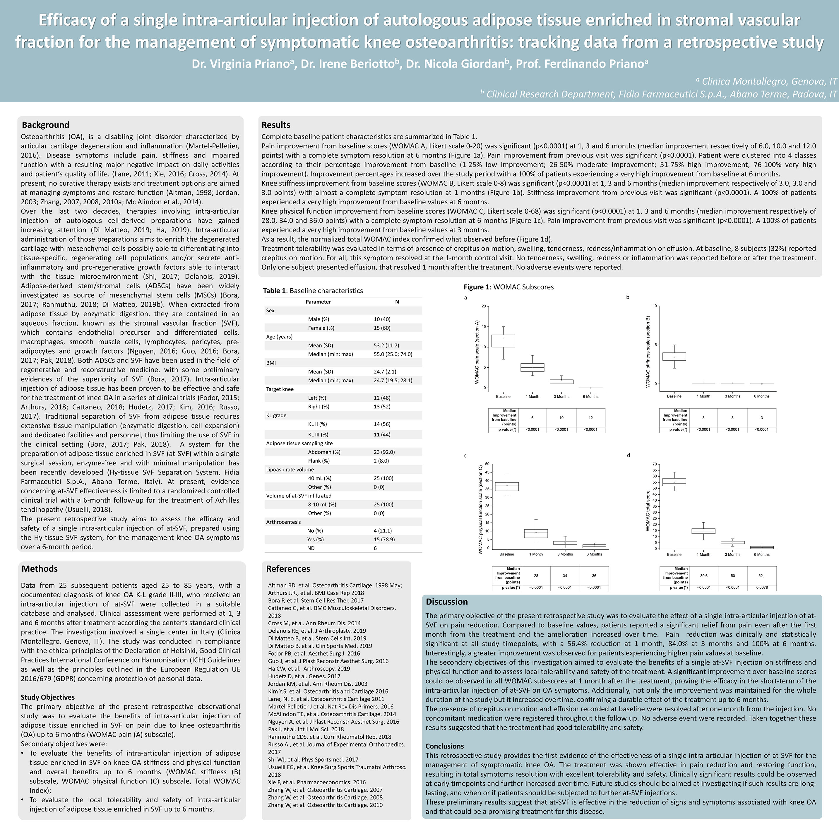
Pain improvement from baseline scores (WOMAC A, Likert scale 0-20) was significant (p<0.0001) at 1, 3 and 6 months (median improvement respectively of 6.0, 10.0 and 12.0 points) with a complete symptom resolution at 6 months (Figure 1a). Pain improvement from previous visit was significant (p<0.0001). Patient were clustered into 4 classes according to their percentage improvement from baseline (1-25% low improvement; 26-50% moderate improvement; 51-75% high improvement; 76-100% very high improvement). Improvement percentages increased over the study period with a 100% of patients experiencing a very high improvement from baseline at 6 months.
Knee stiffness improvement from baseline scores (WOMAC B, Likert scale 0-8) was significant (p<0.0001) at 1, 3 and 6 months (median improvement respectively of 3.0, 3.0 and 3.0 points) with almost a complete symptom resolution at 1 months (Figure 1b). Stiffness improvement from previous visit was significant (p<0.0001). A 100% of patients experienced a very high improvement from baseline values at 6 months.
Knee physical function improvement from baseline scores (WOMAC C, Likert scale 0-68) was significant (p<0.0001) at 1, 3 and 6 months (median improvement respectively of 28.0, 34.0 and 36.0 points) with a complete symptom resolution at 6 months (Figure 1c). Pain improvement from previous visit was significant (p<0.0001). A 100% of patients experienced a very high improvement from baseline values at 3 months.
As a result, the normalized total WOMAC index confirmed what observed before (Figure 1d).
Treatment tolerability was evaluated in terms of presence of crepitus on motion, swelling, tenderness, redness/inflammation or effusion. At baseline, 8 subjects (32%) reported crepitus on motion. For all, this symptom resolved at the 1-month control visit. No tenderness, swelling, redness or inflammation was reported before or after the treatment. Only one subject presented effusion, that resolved 1 month after the treatment. No adverse events were reported.
Discussion
The primary objective of the present retrospective study was to evaluate the effect of a single intra-articular injection of at-SVF on pain reduction. Compared to baseline values, patients reported a significant relief from pain even after the first month from the treatment and the amelioration increased over time. Pain reduction was clinically and statistically significant at all study timepoints, with a 56.4% reduction at 1 month, 84.0% at 3 months and 100% at 6 months. Interestingly, a greater improvement was observed for patients experiencing higher pain values at baseline.
The secondary objectives of this investigation aimed to evaluate the benefits of a single at-SVF injection on stiffness and physical function and to assess local tolerability and safety of the treatment. A significant improvement over baseline scores could be observed in all WOMAC sub-scores at 1 month after the treatment, proving the efficacy in the short-term of the intra-articular injection of at-SVF on OA symptoms. Additionally, not only the improvement was maintained for the whole duration of the study but it increased overtime, confirming a durable effect of the treatment up to 6 months.
The presence of crepitus on motion and effusion recorded at baseline were resolved after one month from the injection. No concomitant medication were registered throughout the follow up. No adverse event were recorded. Taken together these results suggested that the treatment had good tolerability and safety.
Conclusions
This retrospective study provides the first evidence of the effectiveness of a single intra-articular injection of at-SVF for the management of symptomatic knee OA. The treatment was shown effective in pain reduction and restoring function, resulting in total symptoms resolution with excellent tolerability and safety. Clinically significant results could be observed at early timepoints and further increased over time. Future studies should be aimed at investigating if such results are long-lasting, and when or if patients should be subjected to further at-SVF injections.
These preliminary results suggest that at-SVF is effective in the reduction of signs and symptoms associated with knee OA and that could be a promising treatment for this disease.
References
Altman RD, et al. Osteoarthritis Cartilage. 1998 May; Arthurs J.R., et al. BMJ Case Rep 2018
Bora P, et al. Stem Cell Res Ther. 2017
Cattaneo G, et al. BMC Musculoskeletal Disorders. 2018
Cross M, et al. Ann Rheum Dis. 2014
Delanois RE, et al. J Arthroplasty. 2019
Di Matteo B, et al. Stem Cells Int. 2019
Di Matteo B, et al. Clin Sports Med. 2019
Fodor PB, et al. Aesthet Surg J. 2016
Guo J, et al. J Plast Reconstr Aesthet Surg. 2016
Ha CW, et al. Arthroscopy. 2019
Hudetz D, et al. Genes. 2017
Jordan KM, et al. Ann Rheum Dis. 2003
Kim Y.S, et al. Osteoarthritis and Cartilage 2016 Lane, N. E. et al. Osteoarthritis Cartilage 2011 Martel-Pelletier J et al. Nat Rev Dis Primers. 2016 McAlindon TE, et al. Osteoarthritis Cartilage. 2014 Nguyen A, et al. J Plast Reconstr Aesthet Surg. 2016 Pak J, et al. Int J Mol Sci. 2018
Ranmuthu CDS, et al. Curr Rheumatol Rep. 2018 Russo A., et al. Journal of Experimental Orthopaedics. 2017
Shi WJ, et al. Phys Sportsmed. 2017
Usuelli FG, et al. Knee Surg Sports Traumatol Arthrosc. 2018
Xie F, et al. Pharmacoeconomics. 2016
Zhang W, et al. Osteoarthritis Cartilage. 2007
Zhang W, et al. Osteoarthritis Cartilage. 2008
Zhang W, et al. Osteoarthritis Cartilage. 2010


Present with Google Meet
Invite as many as 30 people, and present your poster in high definition. Transcription option is available. Free to use.